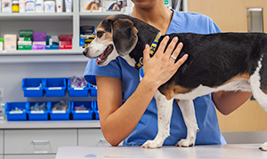
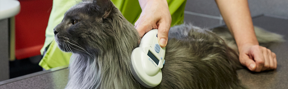
Considering the Epidermal Barrier in the Management of Dermatology Cases
The skin is also an integral part of the innate immune system with the epidermal barrier forming the primary line of defence against the external environment. Far from being an inert structure, this barrier utilises both mechanical and chemical strategies to defend against the constant assault on the skin from external pathogens and allergens. Factors which affect the integrity or function of the epidermis can quickly result in cutaneous infection and/or inflammation; it is therefore crucial that skin barrier abnormality should not just be viewed as a consequence of inflammatory skin disease but also considered as an important, and in some cases primary, triggering factor.
Factors that adversely affect the epidermal barrier are numerous and varied, but it is helpful to consider the structure of the barrier to understand where compromise could occur. The stratum corneum (outermost epidermal layer) is the primary mediator of the epidermal permeability barrier function. A brick wall analogy may be used to describe it, whereby the corneocytes/squames represent the bricks and a lipid intracellular matrix represents the mortar holding the structure together. The lipid matrix consists of ceramides, cholesterol and long chain fatty acids which are arranged in organised lamellar sheets. The healthy stratum corneum provides an effective barrier against potential pathogens and allergens, however it has been demonstrated that atopic dogs appear to lack this highly organised structure, even in skin which appears grossly normal. A study using electron microscopy and ruthenium tetraoxide staining examined skin biopsies from confirmed atopic dogs and compared these to samples from normal dogs; in the atopic animals, corneocytes were vacuolated and the bands of lipids disorganised and thinner1. Many studies in human medicine also suggest that a defective epidermal barrier in combination with an abnormal immune response may contribute to the pathophysiology of atopic dermatitis2.
The surface of the epidermis is covered in a hydrolipd film; this emulsion is formed from apocrine and sebaceous gland secretion which keep the skin supple, regulate TEWL and act as a further microbe barrier. Glandular secretion is under complex hormonal and/or nervous control; apocrine secretions contain a variety of interferons and antibodies, whereas the main component of the oily sebaceous gland secretion is linoleic acid. Endocrine disease, a poor plane of nutrition (particularly with regards to the fatty acid component) and genetic make-up are just some of the factors which can alter the quality, and therefore the function, of this emulsion.
In summary, as medicine continually advances, an ever increasing array of therapeutic options become available for treating dermatological conditions, but the importance of addressing the needs of the epidermal barrier should not be overlooked due to the key roles previously highlighted. As well as allergen avoidance measures, the use of appropriate veterinary shampoos, topical lipid products and diets designed to support dermatological function should be considered alongside anti-inflammatory and/or immunomodulatory drug options when devising treatment strategies for allergic and inflammatory skin disease.
1. Inman AO et al. Electron microscopic observations of stratum corneum intercellular lipids in normal and atopic dogs. Vet Pathol. 2001 Nov;38(6):720-3
2. Lee HJ, Lee SH. Epidermal permeability barrier defects and barrier repair therapy in atopic dermatitis. Allergy Asthma Immunol Res. 2014; 6: 276–87