Oxidative Stress : Its Effect on Cattle and the Role of Trace Minerals
What is Oxidative Stress?
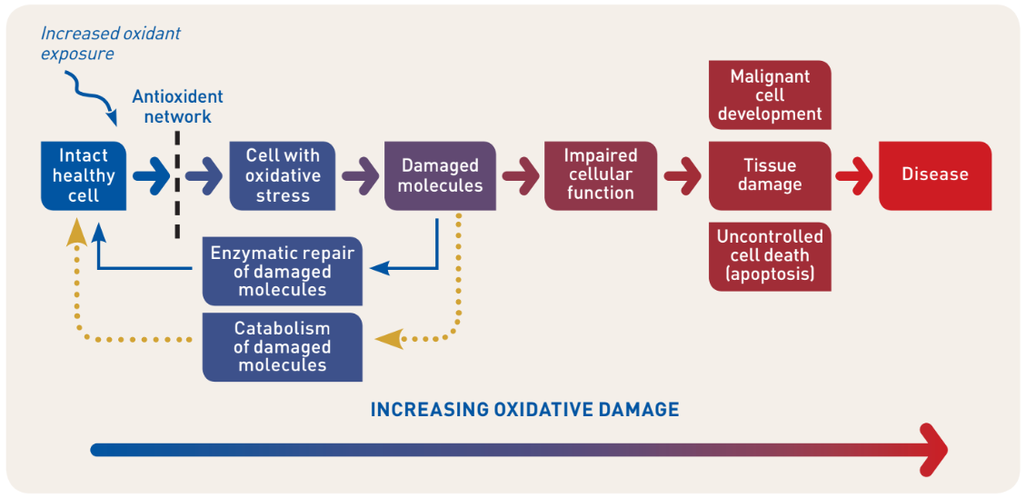
Antioxidants enzymes neutralise free radicals, however, during critical periods, imbalances can result in free radical generation beyond the neutralising capacity of the antioxidants, leading to a disruption of oxido-reduction signalling and control and/or damage to biological molecules such as DNA, proteins and lipids in a process known as Oxidative Stress. (Fig 1)1
Increased oxidative stress is initially counteracted by the host’s antioxidant network. Damaged molecules are either repaired or catabolised and controlled cell suicide (apoptosis) can be initiated if further oxidative damage leads to impaired cellular function.
However, when these signalling cascades are damaged or the oxidative damage exceeds the capacity of the host’s defence mechanisms, uncontrolled cell death, tissue damage and malignant cell development can progress to states of disease2.
Fig.1 Schematic outline of oxidative stress-mediated cellular damage
and progression to disease
Oxidative stress is an imbalance of free radicals and antioxidants in the body, which can lead to cell and tissue damage.
Increased oxidative stress is initially counteracted by the host’s antioxidant network. Damaged molecules are either repaired or catabolised and controlled cell suicide (apoptosis) can be initiated if further oxidative damage leads to impaired cellular function.
However, when these signalling cascades are damaged or the oxidative damage exceeds the capacity of the host’s defence mechanisms, uncontrolled cell death, tissue damage and malignant cell development can progress to states of disease2.
Fig.1 Schematic outline of oxidative stress-mediated cellular damage
and progression to disease
Antioxidants and the Role of Trace Minerals
Free radical production during oxygen metabolism, has necessitated the evolution of highly effective antioxidant defences. These defences can trap reactive intermediates before causing oxidative damage to macromolecules or can reduce biomolecules that have already been oxidised.
Two vital antioxidant metalo-enzyme classes are: the Superoxide Dismutases (SODs), and the family of selenoenzymes, especially Glutathione Peroxidase (GPx)3 .
The structure of bovine SODs and selenoenzymes have been extensively described. Manganese, copper and zinc are key enzymatic cofactors of SOD and selenium is a non-substitutable cofactor of GPx 4 .
The SODs are among the most efficient antioxidants and are considered the first line of defence against oxidative challenge.
Three distinct isoforms of SOD have been identified: two with copper and zinc at their catalytic sites are localised either in the cytoplasm or extracellular, and a third is isoform with manganese as a cofactor localised in the mitochondria.
For the selenium dependent antioxidant enzyme GPx it is clearly acknowledged that this cannot be replaced by any other selenoprotein in protecting against generalised oxidative stress.
Fig 2 shows the role of trace minerals in the reduction of free radicals through antioxidant metalo-enzymes5
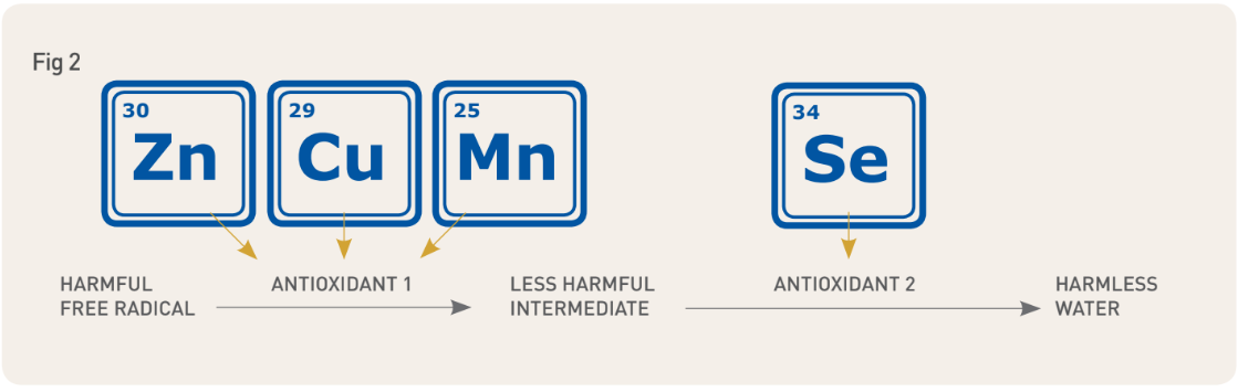
The trace minerals Zinc, Copper Manganese and Selenium are needed for the synthesis of antioxidant metalo-enzymes that neutralise harmful free radicals. Unless there are sufficient available trace minerals to produce these antioxidant metalo-enzymes then animals will suffer from oxidative stress.
The Impact of Oxidative Stress in Cattle
In humans, continued oxidative stress is a known cause of chronic inflammation, which could mediate many chronic diseases including cancer, diabetes, cardiovascular, neurological, and pulmonary diseases6 .
In ruminant medicine oxidative stress has been associated with numerous conditions, including those that are relevant for animal production and the general welfare of the individual animals7 .
In cows, impaired immunological response during oxidative stress periods is suggested by several observations8,9. In weaning calves, transportation stress increases serum concentrations of oxidative stress biomarkers that are related to episodes of BRD and mortality in calves10.
Herds with marginal or defi cient plasma concentrations of Zn or Cu were found to have increased risk of metritis, mastitis, and locomotion problems11. Mastitis challenges have resulted in decreased serum concentrations of Zn and Cu12,13.
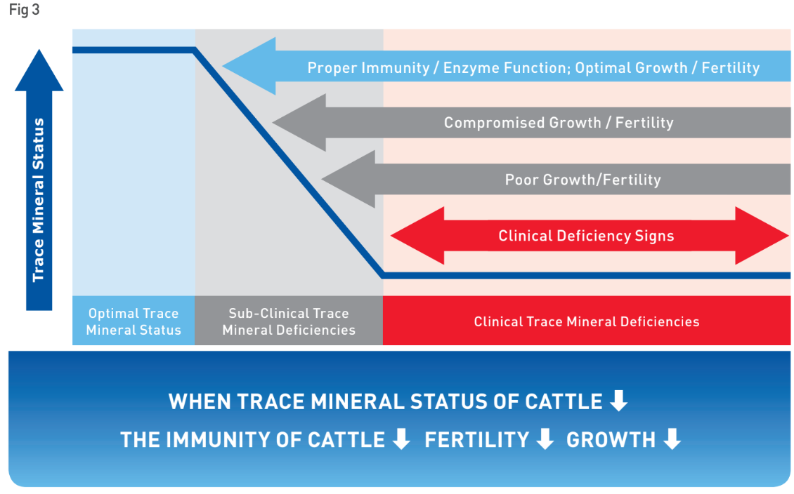
Oxidative stress reduces the health and performance of affected animals.
Indeed when the trace mineral status of cattle declines, immunity and enzyme functions are compromised first, followed by a reduction in growth and fertility and finally a decrease in normal growth prior to clinical deficiency as shown in Fig 3 14.
![large animal graph 5.PNG]() What Factors Might Induce Oxidative Stress?
What Factors Might Induce Oxidative Stress?
 What Factors Might Induce Oxidative Stress?
Significantly, in cattle, high demand periods, which cause accelerated cellular metabolism, such as infection, parturition, drought and heat-stress, as well as embryonic and foetal development, all increase the risk of oxidative stress and as a result, reduced performance 15,16.
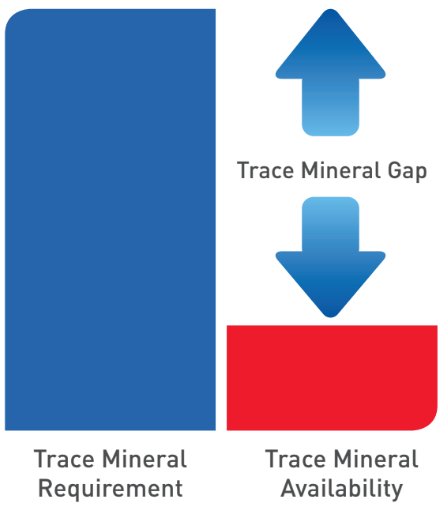
Currently, trace mineral requirements for cattle are primarily provided through their diet and various forms of oral supplementation.
Whilst international guidelines exist in terms of trace mineral feeding levels, reduced oral intake, poor absorption from the rumen and antagonism with other minerals means that during periods of high demand, even in apparently well orally supplemented animals, a trace mineral gap can occur whereby animals are unable to manufacture enough antioxidant enzymes to prevent the negative impacts of oxidative stress.
SUMMARY
Cattle in optimal trace mineral status ahead of high demand periods, are more able to produce the antioxidant enzymes required to combat the increased risk of oxidative stress.
Strategic supplementation ahead of these high demand periods has been shown to mitigate the risk of oxidative stress and improve the health and performance of cattle.17