The Use of Ciclosporin in Sebaceous Adenitis

HISTORY AND CLINICAL EXAM
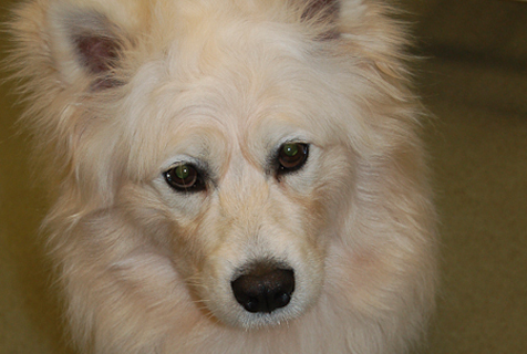
 A 2-year-old, male, neutered, Samoyed dog presented to the clinic with a six-month history of progressive hair loss. There was thinning of his normally thick hair coat was noticed first. This was not initially pruritic, although a mild pruritus developed with time and was present at the time of referral. Over the previous six months, treatment prescribed prior to referral included weekly 2% miconazole and 2% chlorhexidine baths and an oral omega 3 and omega 6 essential fatty acid supplement.
On initial presentation, a full clinical and dermatological examination was performed. The clinical examination was normal. On further dermatological examination, the findings were largely normal although a generalised hypotrichosis and follicular casting was noted. He was up to date with flea and tick prophylaxis, his routine worming and his vaccinations. He had no significant previous health problems. He was fed a raw diet.
DIFFERENTIALS
-
Demodicosis
-
Bacterial folliculitis
-
Sebaceous adenitis
-
Primary seborrhoea
-
Follicular dysplasia
DIAGNOSTICS
To make a definitive diagnosis, it is important to systematically rule out the differential diagnoses for the clinical signs. Skin scrapings and trichograms were performed and were negative for demodex mites. Skin surface cytology was unremarkable. Skin biopsies from affected areas of the coat were obtained and then submitted for dermatohistopathology. This histopathology result confirmed a diagnosis of sebaceous adenitis. The histological findings shown are generally of a reduced number or an absence of sebaceous glands, sparing the hair follicles themselves. During the active phase of the disease, an inflammatory infiltrate can be seen around the sebaceous glands themselves. These findings were observed in this case.
TREATMENT
He was started on 5mg/kg ciclosporin PO SID and an oral omega 3 and omega 6 supplement. The dog did not tolerate the addition of this omega 3 and 6 supplement to the diet. Treatment was initiated with a topical spot on product containing ceramides, fatty acids and cholesterol; used initially weekly, then reduced as the condition improved to once monthly after the first two months of treatment. Three months on, the dose of daily ciclosporin was reduced by 20% and, after a further three months, this dose was administered every other day. This was the result of a 90% improvement in the clinical signs of the dog; it is important to recognise he will never be fully cured, but a 90% improvement is a significant result as the clinical signs are now such that there is an excellent quality of life experienced in this case. He is currently maintained on oral ciclosporin and topical ceramides, fatty acids and cholesterol supplementation.
It was recommended that his diet be changed from a raw diet to a commercially available kibble dog food whilst on ciclosporin therapy. This treatment suppresses the immune system to achieve its results; the bacterial contamination possible in raw food may make him more susceptible to disease and lead to clinical signs, such as diarrhoea. Whilst the vast majority of his coat has recovered, there are still a few patches of hypotrichosis and follicular casting on his limbs and around his head. These areas can be prone to secondary infection; these can be managed with topical antimicrobial agents, such as the use of a chlorhexidine shampoo or topical wipes.
DISCUSSION
Sebaceous adenitis is characterised by an inflammatory insult on the sebaceous glands, resulting in their progressive destruction, leading to classic histopathological changes.1 These changes are diagnostic for the condition and this shows the value of histopathology in the diagnosis of dermatological conditions.
The aetiology is unknown but certain breeds, such as Standard Poodles, Samoyeds and Akitas are predisposed to the condition; it is, however, sporadically seen in other breeds and the Springer Spaniel is over-represented in the UK.2,3 The condition has also been reported in cats and rabbits.
Treatment requires lifelong management and is palliative, with rare cases of complete spontaneous remission.4 Clinical signs may wax and wane irrespective of treatment.5 The aim of treatment is immunotherapy alongside actions to remove excess scale, avoid secondary infection, improve hair coat quality, and restore barrier function of the skin.6 Treatment options include the use of shampoos, both keratolytic and antimicrobial when necessary; emollient rinses; oral supplementation with a omega 3 and omega 6 fatty acid supplementation and / or topical application of ceramides, fatty acids and cholesterol; 50-75% propylene glycol and water sprays; emollient / oil soaks; oral tetracycline and nicotinamide; oral vitamin A; oral synthetic retinoids (isotretinoin and actiretin) and finally oral ciclosporin use for immunomodulation. Glucocorticoids are usually ineffective.7 There have been no controlled studies to document the efficacy of different treatments and individual results may be variable.5
Client education is important due to the variable response to treatment and long term nature of the condition and its management.8 Ciclosporin has been shown to be effective at an oral dosage of 5mg/kg/day; it is the only treatment to show an increase in the numbers of sebaceous glands, possibly due to anagen induction of hair follicles resulting in a removal of excess keratin.8,9 It has been shown it is best used in combination with appropriate topical treatments and was found to cause a decrease in scaling, alopecia and in inflammation of sebaceous glands.9 Affected dogs can be predisposed to secondary skin infections, such as with bacteria or Malassezia spp., due to the defect in the normal skin barrier, and with the reduction of sebum production, due to the destruction of the sebaceous glands. An awareness of this is important in the management of these cases and to ensure owner compliance in the treatment.